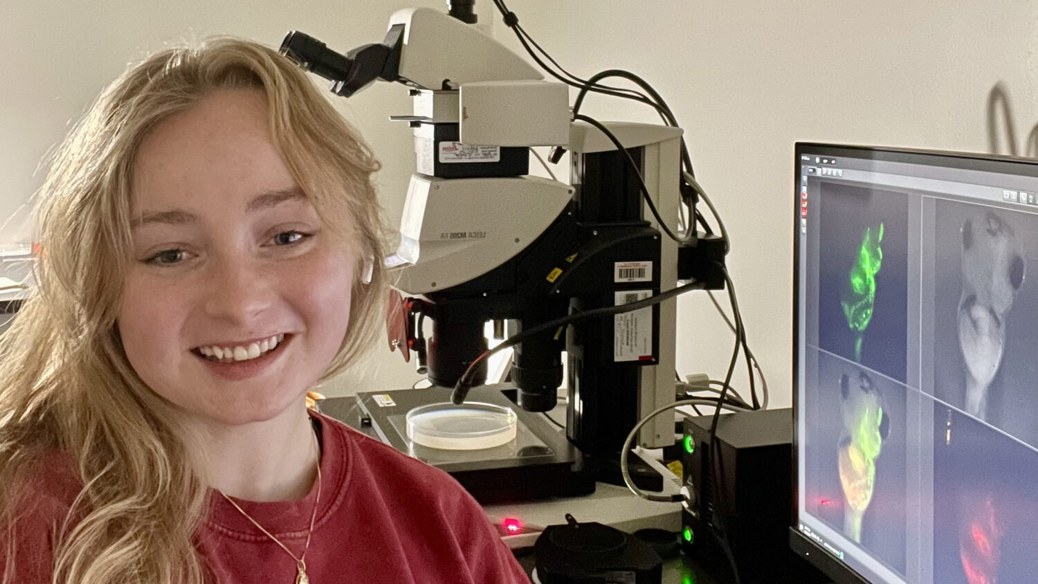
I’m a fifth-year PhD candidate in the Genetics and Genomics program at NC State. I work in Nanette Nascone-Yoder’s lab at the College of Veterinary Medicine, where we study left-right asymmetry, a highly conserved and fundamental feature of our internal anatomy. We use Xenopus laevis frog embryos to study how left-right asymmetry develops in the individual organs of the digestive tract. Their translucent skin allows us to observe gut development that is remarkably similar to that of humans, and genetic manipulations to this process can be targeted to individual organs via microinjection at early blastula stages. I’m passionate about mentoring students interested in research and exploring new hypotheses. My long-term career goals involve pursuing a position as research faculty or perhaps as an imaging facility director. Outside of the lab, I love taking my dog to kayak on Lake Raleigh or bike on the greenway, hanging out with friends, playing volleyball, or reading a book.
What instruments are you using for your research and why do you like them?
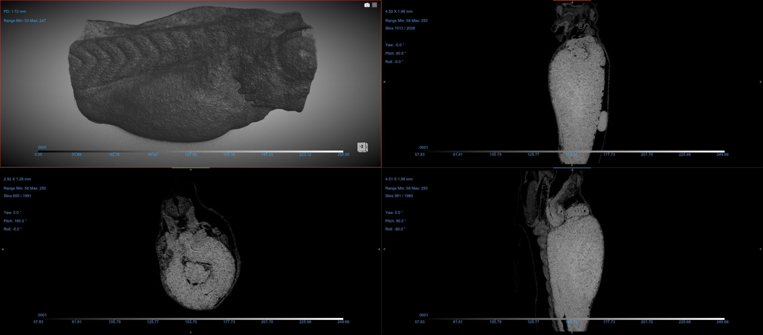
As the gut’s left-right asymmetry develops, complex tissue rearrangements occur across multiple spatial dimensions and cellular layers. Traditionally, we’ve relied on labor-intensive histological sectioning to reveal internal tissue architecture in a limited 2D format. Using the AIF’s Zeiss Xradia 510 Versa 3D X-ray Tomography System (nano-CT) has allowed me to generate high-resolution 3D data quickly and reliably. The Xradia nano-CT enables rapid assessment of internal tissue architecture in whole Xenopus embryos without time-consuming sectioning, while optical sections can reveal the exact 2D/3D geometries of left and right stomach tissues. Segmenting individual organs and tissue layers on the AIF’s CT data analysis workstation allows us to generate 3D meshes that our collaborators use to create cellularized 3D models directly comparable to in vivo morphologies. This integrated approach enables limitless in silico exploration of morphogenetic mechanisms while maintaining quantitative comparisons to real organ geometry.
What have you been researching?
My dissertation project explores the genetic and developmental mechanisms underlying left-right asymmetric organogenesis, with a particular focus on extracellular matrix (ECM) dynamics. Specifically, I’m investigating how the fibronectin matrix regulates stomach curvature through its influence on tissue stiffness, compaction, adhesion, and surface tension. To elucidate these complex interactions, I’m using microinjection and microsurgery to inhibit fibronectin translation and/or binding. I evaluate the effect this has on gut morphology via immunohistochemistry, atomic force microscopy, and nano-CT, complemented by computational modeling.
My research aims to uncover fundamental genetic and biomechanical principles governing left-right asymmetric organogenesis and their implications for serious laterality-related birth defects. Understanding these embryonic events is vital for determining the causes of heterotaxy: a syndrome occurring in 1 in every 10,000 births when the left-right axis is not properly established during development, resulting in organs with abnormal morphology or that are missing entirely. Children with heterotaxy often undergo multiple complex surgeries, and the disorder carries a devastating one-year mortality rate of up to 85%. Left-right anomalies in individual organs, such as congenital heart defects and intestinal malrotations, occur at even higher frequencies. Despite their prevalence, the genetic and molecular etiology of these conditions remains largely unknown. By understanding how left-right asymmetry develops in individual organs, we can better determine the causes of heterotaxy and other common laterality-related birth defects.
What have you learned from your experience at AIF?
I’ve learned that AIF offers many valuable resources and opportunities. I’ve enjoyed classes hosted at the AIF, namely BIT 495 Introduction to Electron Microscopy, and appreciated the access to instrumentation that has positively contributed to my thesis and research experience at NC State. Most specifically, I’ve learned extensively about micro-CT, how it works, and how to best analyze the data it produces.
“Using AIF’s nano-CT system has transformed how I study gut development, allowing me to visualize complex 3D tissue structures that were once only possible through time-consuming 2D methods.”
– Carley Huffstetler
Best thing about AIF in 5 words or less?
Accessible cutting-edge instruments, insightful staff.
Is there a staff member at AIF that has helped you?
Ruksana Baby has been extremely supportive of my work with the X-Radia nano-CT, providing training and kindly answering my questions since the first year of my PhD. She is always helpful, knowledgeable, and communicative.
I’m also grateful to AIF’s Aaron Bell, who taught my BIT Electron Microscopy class. He got me excited about electron microscopy and went out of his way to help me obtain some useful and interesting preliminary data during the course. Both Ruksana and Aaron have greatly influenced my career goals!
This post was originally published in Analytical Instrumentation Facility (AIF).
- Categories: